


Abdominal MRA
Two-In-One Imaging
Gadovist®는 한번의 검사에서 복부 소혈관(MRA)과 장기 실질(MRI)의 영상 획득을 가능하게 합니다.
두 가지 검사를 하나로 결합하면 스캔에 필요한 조영제의 양이 줄어들어 검사 과정과 비용이 더 효율적으로 되고 환자는 더 편해집니다.7


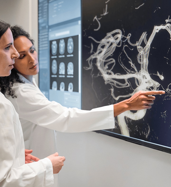
Supra-Aortic MRA
High Quality Static and Dynamic Imaging
Gadovist®는 높은 SNR과 CNR을 모두 가능하게 함으로써 우수한 품질의 영상을 제공할 수 있습니다.
20명의 건강한 지원자를 대상으로 한 개체 내 비교 연구에서 Gadovist®는 0.5몰 거대고리형 가도테레이트 메글루민보다 근위 내경동맥(ICA) 수준에서 더 높은 SNR을 보여주었습니다(p < 0.05).8
MRA vs DSA
The Less Invasive Alternative


References
- 1. Szomolanyi et al. Comparison of the Relaxivities of Macrocyclic Gadolinium-Based Contrast Agents in Human Plasma at 1.5, 3, and 7 T, and Blood at 3 T. Invest Radiol. 2019;54(9):559-564. Return to content
- 2. Shen et al. T1 relaxivities of gadolinium-based magnetic resonance contrast agents in human whole blood at 1.5, 3, and 7 T. Invest Radiol. 2015;50(5):330-338. Return to content
- 3. Rohrer et al. Comparison of magnetic properties of MRI contrast media solutions at different magnetic field strengths. Invest Radiol. 2005;40(11):715-724. Return to content
- 4. Tombach et al. Comparison of 1.0 M gadobutrol and 0.5 M gadopentate dimeglumine-enhanced MRI in 471 patients with known or suspected renal lesions: results of a multicenter, single-blind, interindividual, randomized clinical phase III trial. Eur Radiol. 2008;18(11):2610-2619. Return to content
- 5. Tombach B, Heindel W. Value of 1.0- M gadolinium chelates: review of preclinical and clinical data on gadobutrol. Eur Radiol. 2002;12(6):1550-1556. Return to content
- 6. Gutierrez et al. Safety and Efficacy of Gadobutrol for Contrast-enhanced Magnetic Resonance Imaging of the Central Nervous System: Results from a Multicenter, Double-blind, Randomized, Comparator Study [published correction appears in Magn Reson Insights. 2015;8:23]. Magn Reson Insights. 2015;8:1-10. Return to content
- 7. Liu et al. Improved display of abdominal contrast-enhanced MRA using gadobutrol: comparison with Gd-DTPA. Clin Radiol. 2019;74(12):978.e1-978.e7. Return to content
- 8. Kramer et al. Dynamic and static magnetic resonance angiography of the supra-aortic vessels at 3.0 T: intraindividual comparison of gadobutrol, gadobenate dimeglumine, and gadoterate meglumine at equimolar dose. Invest Radiol. 2013;48(3):121-128. Return to content
- 9. Hadizadeh et al. Noninvasive evaluation of cerebral arteriovenous malformations by 4D-MRA for preoperative planning and postoperative follow-up in 56 patients: comparison with DSA and intraoperative findings. AJNR Am J Neuroradiol. 2012;33(6):1095-1101. Return to content
- 10. Kukuk et al. Cerebral arteriovenous malformations at 3.0 T: intraindividual comparative study of 4D-MRA in combination with selective arterial spin labeling and digital subtraction angiography. Invest Radiol. 2010;45(3):126-132. Return to content
- 11. Burbelko et al. Comparison of contrast-enhanced multi-station MR angiography and digital subtraction angiography of the lower extremity arterial disease. J Magn Reson Imaging. 2013;37(6):1427-1435. Return to content
- 12. Budjan et al. Diffusion kurtosis imaging of the liver at 3 Tesla: in vivo comparison to standard diffusion-weighted imaging. Acta Radiol. 2018;59(1):18-25. Return to content